


Echocardiography applies harmless sound waves influentially and rapidly to reach precious information about your heart. The valuable information may include heart performance, size and shape. It is a noninvasive versatile tool to help to evaluate heart valves and detect heart conditions. It can be done through different methods depending on your heart problem.
What is echocardiography?
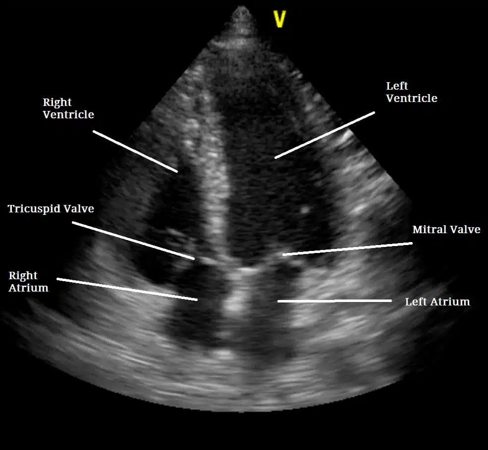
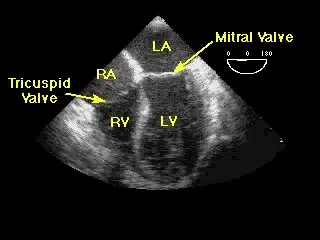
Echocardiography, also known as echo and echocardiogram, is a painless test used to diagnose heart abnormalities such as damage to the heart tissue from heart attack or poorly functioning heart valves. Echocardiography uses ultrasound waves to visualize the heart. These waves echo off the heart. They can display the shape, size and motion of the heart’s atria, ventricles and valves. Echocardiography can also show the flow of blood through the heart.
What is the purpose of echocardiography?
This diagnostic test is used to find probable certain cardiovascular diseases. Echocardiography is one of the most widely used tests to diagnose heart disease. Echocardiography can provide plenty of helpful data, including the size, and shape of your heart, the location and extent of any damage to the heart’s tissues and its pumping strength.
Echocardiogram Description
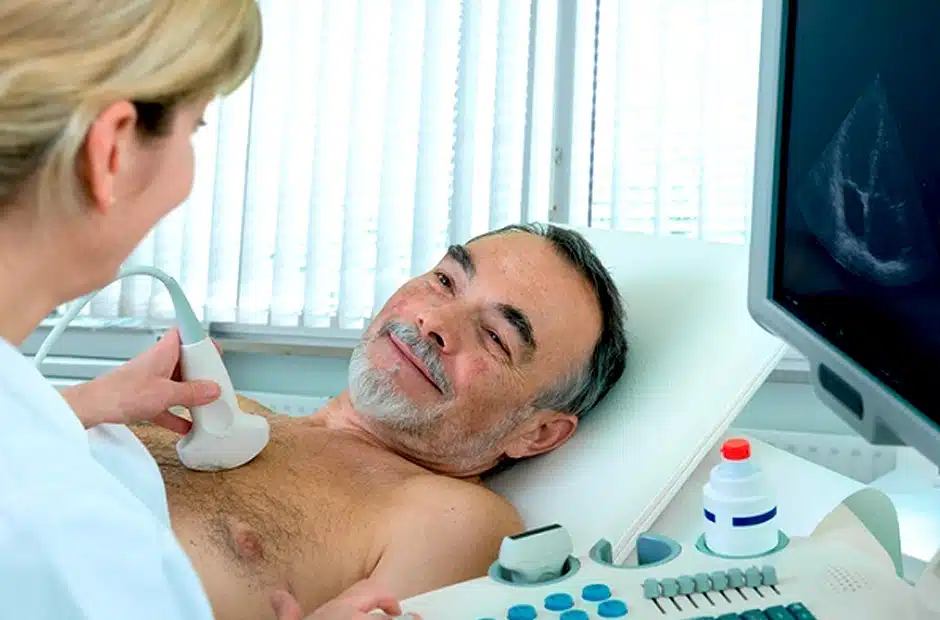
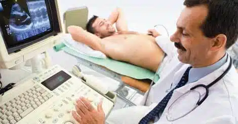
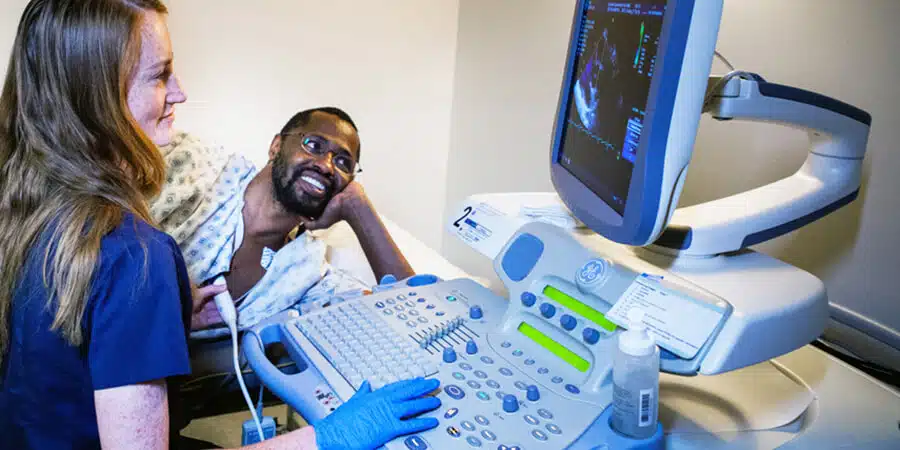
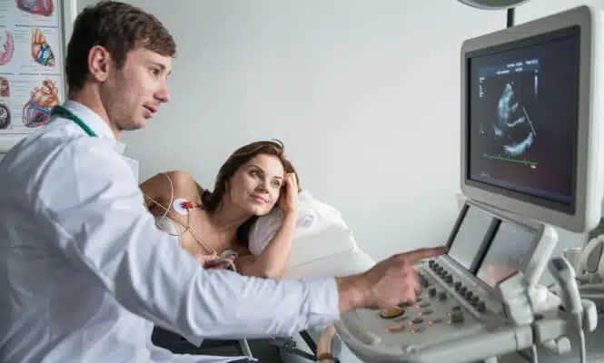
Transthoracic echocardiogram method (TTE)
This method is similar to ultrasound scanning that is commonly used during the pregnancy to visualize the fetus. You lie bare chested on an examination table and the radiologist spreads a special gel over your chest to help the transducer slide smoothly over the skin and make good contact. The doctor places a transducer, a small hand-held device, against your chest. It directs ultrasound waves into the chest. Some of the waves reflect (echoed) to the device.
3Dimentional echocardiography
This method typically displays a 3_D picture as well as a flat picture. It is specifically helpful to detect problems with heart valves and lower left chambers.
Intracardiac echocardiogram (ICE)
This method displays a newer form of testing with images that are taken inside the heart. ICE is mostly used to monitor treatments that involve the placement of thin tubes (catheters) inside the heart arteries.
Stress echocardiography
This method is applied as a part of an extensive stress test that intentionally increases the heart rate and blood pressure. Two sets of images are taken, one at working out on a treadmill or stationary bike and another at rest. If your heart condition prevents the exercise, you will be prescribed motivating medicines which is called pharmacologic echocardiography.
Trans-esophageal echocardiogram
In this approach a special ultrasound probe is guided into the patient’s mouth and down his/her esophagus after adequately sedation. Then, the doctors can get better images, as the heart and the esophagus sites are close to each other and the waves of the ultrasound are not required to pass through muscle, skin or bone.
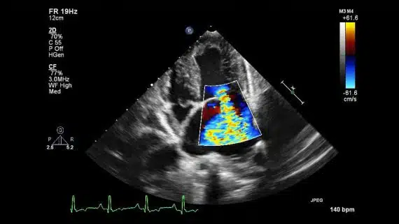
Doppler echocardiography
This method is used to evaluate and measure the blood flow through the valves and chambers of the heart. The amount of blood that pumps out with each beat is the indicator of the heart’s functioning. Doppler echocardiography can also detect blood flood abnormalities within the heart septum.